Glucose Dehydrogenase (FADGDH-AD) 葡萄糖脱氢酶

  • 临床检测用酶
葡萄糖脱氢酶

FADGDH-AD is an FAD-dependent glucose dehydrogenase with low reactivity toward maltose and xylose. It is one of the most stable glucose dehydrogenases for blood glucose measurement and is suitable for continuous glucose monitoring sensor.

由来 recombinant Aspergillus sojae
系统名称

D-Glucose : acceptor 1-oxidoreductase

EC编号 1.1.5.9
反应式

D-Glucose + acceptor →→→ D-Glucono-1,5-lactone + reduced acceptor

规格

Appearance yellow to brown lyophilizate
Activity ≧700 U/mg
Contaminants NAD glucose dehydrogenase  <0.01 U/U%
Hexokinase  <0.01 U/U%
α-glucosidase  <0.01 U/U%
β-glucosidase  <0.01 U/U%
Storage condition below -20℃ protected from light

特性

Molecular weight ca. 90 kDa (SDS-PAGE)
Structure monomer, one mole of FAD per mole of enzyme glycoprotein
Michaelis constant 6.4×10⁻²M (D-glucose)
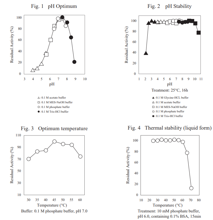
pH Optimum 7.0–7.5 (Fig.1)
pH Stability 2.5–9.5 (Fig.2)
Optimum temperature 45℃ (Fig.3)
Thermal stability (liquid form) below 60℃ (Fig.4)
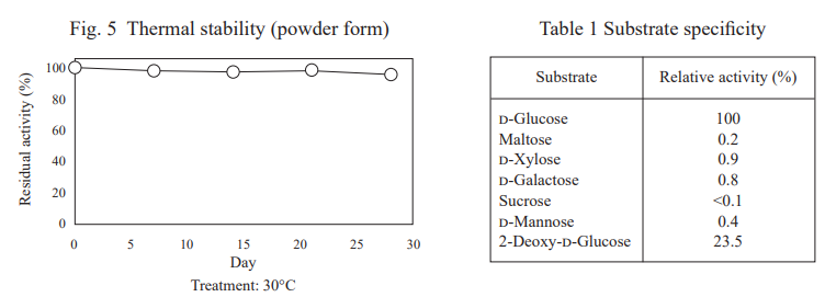
Thermal stability (powder form) stable at 30℃ for at least one month (Fig.5)
Inhibitors Mn2+, Ag+
Specificity (Table.1) D-glucose (100%), maltose (0.2%), 
D-xylose (0.9%), D-galactose (0.8%) 
sucrose (<0.1%),  D-mannose (0.4%)
2-deoxy-D-glucose (23.5%)

适用类型

By using FADGDH-AD and an electron mediator-modified polymer, glucose can be continuously measured by an electrode method.

ASSAY PROCEDURE

Principle

The disappearance of the blue color of DCIP by the reduction is measured spectrophotometrically at 600 nm.

Definition of unit

One unit (U) causes the reduction of one micromole of DCIP per minute under the conditions described below.

Reagents

  1. D-Glucose solution, 2 M: 72 g of d-glucose/200 mL of distilled water.
  2. Potassium phosphate buffer, 0.1 M; pH 7.0: mix 0.1 M KH2PO4 solution and 0.1 M K2HPO4 solution to make a pH 7.0 solution.
  3. 2,6-Dichloroindophenol (DCIP) solution, 1.8 mM: 58.7 mg of DCIP/100 mL of distilled water.
  4. 5-Methylphenazinium methyl sulfate (PMS) solution, 30 mM: 91.9 mg of PMS/10 mL of distilled water.
  5. Enzyme dilution buffer: 10 mM potassium phosphate buffer, pH 6.0, containing 0.1% bovine serum albumin (BSA).

Sample: dissolve the lyophilized enzyme to final concentration about 0.4 μg/mL with enzyme dilution buffer (Reagent E) immediately before measurement.

Procedure

  1. Pipette the following reagents into a cuvette (light path: 1 cm).
       600 μL      D-Glucose solution                            (Reagent A)
       2050 μL    Potassium phosphate buffer pH 7.0  (Reagent B)
       150 μL      DCIP solution                                     (Reagent C)
  2. Equilibrate at 37℃  for about 3 min.
  3. Add 0.1 mL of PMS solution (Reagent D) and mix.
  4. Add 0.1 mL of sample and mix.
  5. Record the decrease of absorbance at 600 nm against water for 1 min. (30–90 sec) in a spectrophotometer thermostated at 37℃ , and calculate the ΔA per min using the linear portion of the curve (ΔAS). The blank solution is prepared by adding Enzyme dilution buffer (Reagent E) instead of sample (ΔA0).

Calculation

Activity can be calculated by using the following formula:

20.4 : Millimolar extinction coefficient of DCIP under the assay condition (cm2 /µmol)
1.0 : Light pass length (cm)
df : Dilution factor

EXPERIMENTAL DATA

Line-up

参考文献

Satake R, Ichiyanagi A, Ichikawa K, Hirokawa K, Araki Y, Yoshimura T, Gomi K (2015)
Novel glucose dehydrogenase from Mucor prainii: Purification, characterization, molecular cloning and gene expression in Aspergillus sojae
Biosci Bioeng., 120, 498-503

Masakari Y, Hara C, Araki Y, Gomi K, Ito K (2020)
Improvement in the thermal stability of Mucor prainii-derived FAD-dependent glucose dehydrogenase via protein chimerization 
Enzyme Microb Technol., 132, 109387, doi: 10.1016/j.enzmictec.2019.109387.
https://doi.org/10.1016/j.enzmictec.2019.109387, (cited 2020-07-02)


TOP